Peri-implantitis is a disease of the tissues around an implant, marked by plaque-related inflammation and accompanying progressive bone loss. Its early stage, peri-implant mucositis, can be reversed with appropriate treatment and care. That is why noticing changes around an implant early and attending regular check-ups are so important.
What is peri-implantitis?
Peri-implantitis is a peri-implant disease defined by inflammation in the soft tissue surrounding the implant and progressive loss of the bone supporting it. It was defined as a separate diagnosis in the classification of periodontal and peri-implant diseases published in 2017.
The main cause of the disease is bacterial plaque that builds up on and around the implant surface. Although it follows a process similar to periodontitis around natural teeth, the structure of the tissues around an implant is different, so in some cases the disease can progress more quickly.
A natural tooth has a connective tissue between the root and the bone (the periodontal ligament) that suspends the tooth and contributes to its defenses. An implant has no such structure; it is in direct contact with the bone, and the gum fibers around it are arranged differently. This difference is considered one of the reasons the tissues around an implant may be more vulnerable to bacterial plaque.
What is the difference between peri-implant mucositis and peri-implantitis?
The key difference is whether there is bone loss. Peri-implant mucositis is inflammation confined to the soft tissue around the implant; there is no bone loss, and it can be reversed with appropriate treatment. Peri-implantitis is the stage at which the inflammation spreads to the bone and causes loss of the bone supporting the implant.
- Peri-implant mucositis: Bleeding on gentle probing, redness and swelling are seen; X-rays show no bone loss beyond what is expected after initial healing.
- Peri-implantitis: In addition to bleeding and/or pus, increased pocket depth and progressive bone loss on X-rays are found.
Leaving mucositis untreated is considered an important step toward peri-implantitis. This is why intervening at an early stage is the most effective step in treatment.
What are the signs of peri-implantitis?
There may be no noticeable complaints at first; for this reason, the disease is often detected during check-ups. The following signs deserve attention:
- Bleeding around the implant during brushing or on its own
- Redness, swelling or tenderness of the gum
- Pus discharge from around the implant
- A bad taste or odor in the mouth
- Gum recession around the implant, with the metal surface becoming visible
- Looseness of the implant at an advanced stage
If you notice any of these signs, see your dentist to have your implant evaluated. The absence of pain does not mean the absence of disease.
What factors increase the risk of peri-implantitis?
Research shows that certain factors increase the likelihood of developing peri-implantitis:
- A history of periodontitis
- Inadequate oral hygiene and not attending regular check-ups
- Smoking
- Uncontrolled diabetes
- A restoration design that makes cleaning difficult, or excess cement left below the gum line
- Insufficient attached gum tissue around the implant
Some of these factors can be changed. For more on the effects of smoking, you can read the article on smoking and gum disease.
How is peri-implantitis diagnosed?
The diagnosis is made by evaluating clinical examination and X-ray findings together. During the examination, the pocket depth around the implant is measured with a fine probe, and bleeding and discharge are checked. On X-rays, the bone level around the implant is compared with X-rays taken when the restoration was placed.
Probing is done with a light force that does not harm the tissues around the implant and is part of the routine check-ups that follow implant treatment. If there is no baseline X-ray or measurement from the time the restoration was placed, the evaluation is based on current findings and the expected bone level of the implant. That is why keeping records after implant treatment and attending regular check-ups make it easier to detect changes early.
The fit of the restoration, how easy it is to clean and how biting forces are distributed are also evaluated. When needed, removing the restoration to examine the area around the implant in more detail may be planned.
How is nonsurgical treatment carried out?
The first step of treatment is nonsurgical therapy. The S3-level clinical practice guideline published in 2023 by the European Federation of Periodontology (EFP) also recommends a stepwise approach. This stage usually includes the following steps:
- Personalized oral hygiene instruction and control of risk factors
- Removal of plaque and tartar from the implant surface with suitable instruments
- Cleaning the area with antiseptic solutions when considered necessary
- Adjusting parts of the restoration that make cleaning difficult and removing any excess cement
The procedure can be performed under local anesthesia; the aim is for you not to feel pain during the procedure. Scientific evidence on the added benefit of adjunctive methods such as lasers is still limited; these methods may be added to treatment based on the clinician's assessment. Peri-implant mucositis can often be brought under control at this step.
A few weeks to a few months after nonsurgical treatment, the tissues around the implant are measured again. If the inflammation has not subsided or deep pockets persist, the surgical step of treatment is planned. This interim reassessment is important for avoiding unnecessary surgery and intervening at the right time.
When is surgical treatment needed for peri-implantitis?
If bleeding, discharge and deep pockets persist despite nonsurgical treatment, surgical treatment is considered. In surgical treatment, the gum is opened to gain direct access to the implant surface, which is then carefully cleaned. Different approaches may be chosen depending on the shape of the bone loss:
- Open flap debridement: Cleaning the area under direct vision and repositioning the tissues.
- Resective approach: Reshaping areas that are difficult to clean so that the site is easier to care for.
- Regenerative approach: In bone defects of a suitable shape, attempting to increase bone support using graft material and, if needed, a membrane.
For implants with advanced bone loss that have become loose, removal of the implant may be necessary. In that case, whether a new implant can be placed after the area heals is evaluated separately. After surgery, swelling, tenderness and some recession of the gum level may occur.
In the first few days after surgery, cold compresses, a soft diet and use of the mouth rinses your dentist recommends are advised. Until the stitches are removed, the area is cleaned gently. Because smoking can impair healing, it is important to avoid smoking during this period.
Why is maintenance after treatment important?
The long-term success of peri-implantitis treatment depends on regular maintenance therapy. Maintenance includes check-ups at set intervals, professional cleaning and a review of your oral care routine. The interval is set according to each person's risk profile and, for most patients, is planned every 3–6 months.
At maintenance appointments, pocket depth and bleeding around the implant are measured again, X-rays are taken when needed, and accumulated plaque is professionally removed. Patients who do not attend regular maintenance are reported to have a higher likelihood of the disease coming back.
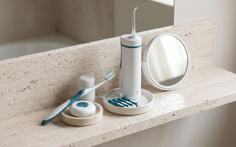
Regular brushing at home and the use of interdental brushes and, if needed, a water flosser are an integral part of treatment. For detailed recommendations, see the article how to care for dental implants. Periodontist Dr. Nadin Gemrekoğlu, DDS, PhD, regards the principles of nonsurgical periodontal therapy, which she also examined in her doctoral research, as the foundation of a stepwise approach to managing peri-implant diseases. If a new implant is being planned, the pre-treatment assessment steps on the dental implants page may also be helpful.